The anatomy of the nervous system : from the standpoint of development and function / by Stephen Walter Ranson.
- Ranson, Stephen Walter, 1880-1942.
- Date:
- 1923
Licence: In copyright
Credit: The anatomy of the nervous system : from the standpoint of development and function / by Stephen Walter Ranson. Source: Wellcome Collection.
35/426 (page 35)
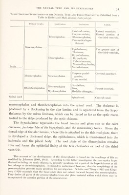
![Table Showing Subdivisions of the Neural Tube and Their Derivatives (Modified from a Table in Keibel and Mall, Human Embryology). Primary vehicles. Subdivisions. Derivatives. Lumen. [ | Prosencephalon... . < i 1 Telencephalon. [ Cerebral cortex, Corpora striata, Rhinencephalon, Pars-optica hypo¬ thalami. Lateral ventricles. Rostral portion of the third ventricle. Brain.! [ Diencephalon.1 Epithalamus, Thalamus, Hypothalamus, Hypophysis, Tuber cinereum, Mammillary bodies, Metathalamus. The greater part of the third ventricle. Mesencephalon { Mesencephalon . . .. ] Corpora quadri- gemina, Crura cerebri. Cerebral aqueduct. 1 f Rhombencephalon Metencephalon j Myelencephalon Cerebellum, Pons, Medulla oblongata.] Fourth ventricle. Spinal cord Spinal cord. Central canal. mesencephalon and rhombencephalon into the spinal cord. The thalamus is produced by a thickening in the alar lamina and is separated from the hypo¬ thalamus by the sulcus limitans, which can be traced as far as the optic recess rostral to the ridge produced by the optic chiasma. The hypothalamus represents the basal lamina and gives rise to the tuber cinereum, posterior lohe of the hypophysis, and the mammillary bodies. From the dorsal edge of the alar lamina, where this is attached to the thin roof plate, there is developed a thickened ridge, the epithalamus, which is transformed into the habenula and the pineal body. The roof plate of the diencephalon remains thin and forms the epithelial lining of the tela chorioidea or roof of the third ventricle. This account of the development of the diencephalon is based on the teachings of His as modified by Johnston (1909, 1912). According to the latter investigator the pars optica hypo¬ thalami including the optic chiasma is, properly speaking, not a part of the hypothalamus at all, but belongs to the telencephalon. It should be understood, however, that the morphology of the prosencephalon presents many problems for investigation. Schulte and Tilney (1915) and Kings¬ bury (1920) maintain that the basal plate does not extend forward beyond the mesencephalon. They derive all parts of the prosencephalon from alar plate material within which there may be incorporated the anterior portion of the neural crest.](https://iiif.wellcomecollection.org/image/b29813669_0035.jp2/full/800%2C/0/default.jpg)