Volume 1
On the anatomy, physiology, and pathology of the chimpanzee / by Charles F. Sonntag.
- Sonntag, Charles F. (Charles Frederick), -1925.
- Date:
- 1923
Licence: In copyright
Credit: On the anatomy, physiology, and pathology of the chimpanzee / by Charles F. Sonntag. Source: Wellcome Collection.
75/118 (page 395)
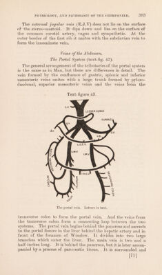
![in the popliteal space and enters the femoral vein. There is no upward vein running through a saphenous opening, and that opening is a human characteristic. Two veme comites accompany all the branches of the posterior tibial artery. They unite to form one popliteal vein which accompanies the artery and becomes the femoral vein. The venous circulation closely follows the arterial supply, but no epigastric vein enters the femoral. The saphenous veins open, as described above, into the popliteal and femoral veins. The veins of the pelvis follow the branches of the hypogastric artery, and the hypogastric vein joins with the external iliac vein to join the common iliac vein. The two common iliac veins unite to form the vena cava inferior. These veins have rela- tions similar to those in Man. Veins of the Pectoral Extremity. The venous circulation differs in several points from that in Man. The veins of the hand unite to form the cephalic vein wdiich only extends up as far as the antecubital fossa. There it dips inwards and unites with veme comites following the branches of the brachial artery to form the brachial vein. No basilic vein is present. The brachial vein runs upwards, re- ceiving tributaries corresponding to the branches of the artery. It is successively followed by the axillary and subclavian veins which receive tributaries corresponding to the branches of the arteries. The subclavian veins unite with the external jugular veins to form the innominate veins. The venous circulation differs from that of Man in the shortness of the cephalic vein, the absence of the basilic vein, the presence of a brachial vein instead of venae comites, and the absence of an internal jugular vein uniting with the innominate vein. The Ductless Glands. The thyroid gland (text-fig. 41) is long, narrow, and thin. The lateral lobes are bent on themselves at the upper ends, which lie against the cricoid and lower end of the thyroid cartilage. The thicker isthmus crosses the fourth and fifth tracheal rings. There is no strong capsule and no pyramidal lobe. It receives a complicated series of arterial anastomoses from the superior (S.T.A), middle (M.T.A), and inferior (I.T.A) branches of the external and common carotids, and the thyroidea ima (T.I.A) from the left common carotid. No subclavian branches pass to the gland. The superior thyroid vein (S.T.V) opens into the anterior facial vein, and the inferior thyroid vein (I.T.V) goes to the innominate vein. At the lower border of the isthmus there is, on each side, an oval body, the size of a pea, consisting of the parathyroid gland and a piece of thymus. No other parathyroid tissue was present. [73]](https://iiif.wellcomecollection.org/image/b2982123x_0001_0075.jp2/full/800%2C/0/default.jpg)