A text-book of pathological anatomy and pathogenesis / by Ernst Ziegler ; tr. and ed. for English students by Donald MacAlister.
- Ernst Ziegler
- Date:
- 1887
Licence: Public Domain Mark
Credit: A text-book of pathological anatomy and pathogenesis / by Ernst Ziegler ; tr. and ed. for English students by Donald MacAlister. Source: Wellcome Collection.
Provider: This material has been provided by the Francis A. Countway Library of Medicine, through the Medical Heritage Library. The original may be consulted at the Francis A. Countway Library of Medicine, Harvard Medical School.
360/1132 (page 326)
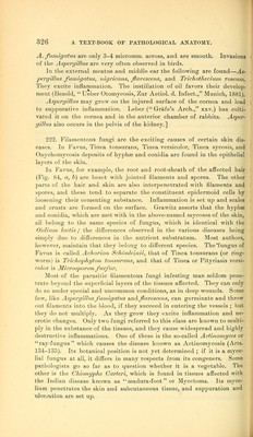
![A. fumigatus are only 3-4 micromm. across, and are smooth. Invasions of the Aspergillus are very often observed in birds. In the external meatus and middle ear the following are found—'^s- pergillus fwnigatics, nigricans, flavescens, and Trichothecium roseum. They excite inflammation. The instillation of oil favors their develop- ment (Bezold, Ueber Otomycosis, Zur Aetiol. d. Infect., Munich, 1881). Aspergillus may grow on the injured surface of the cornea and lead to suppurative inflammation. Leber ( Grafe's Arch., xxv.) has culti- vated it on the cornea and in the anterior chamber of rabbits. Asper- gillus also occurs in the pelvis of the kidney.] 222. Filamentous fungi are the exciting causes of certain skin dis- eases. In Favus, Tinea tonsurans. Tinea versicolor, Tinea sycosis, and Onychomycosis deposits of hyphffi and conidia are found in the epithelial layers of the skin. In Favus, for example, the root and root-sheath of the affected hair (Fig. 84, a, h) are beset with jointed filaments and spores. The other parts of the hair and skin are also interpenetrated with filaments and spores, and these tend to separate the constituent epidermoid cells by loosening their cementing substance. Inflammation is set up and scales and crusts are formed on the surface. Grawitz asserts that the hyphse and conidia, which are met with in the above-named mycoses of the skin, all belong to the same species of fungus, wliich is identical with the O'ldlimi lactis^' the differences observed in the various diseases being simply due to differences in the nutrient substratum. Most authors, however, maintain that they belong to different species. The *fungus of Favus is called Achorion Scho7ileinii, that of Tinea tonsurans (or ring- worm) is Trichophyton tonsurans, and that of Tinea or Pityriasis versi- color is Microsporon furfur. Most of the parasitic filamentous fungi infesting man seldom pene- trate beyond the superficial layers of the tissues affected. They can only do so under special and uncommon conditions, as in deej3 wounds. Some few, like Aspergillus fumigatus and favescens, can germinate and throw- out filaments into the blood, if they succeed in entering the vessels ; but they do not multiply. As they grow they excite inflammation and ne- crotic changes. Only two fungi referred to this class are known to multi- ply in the substance of the tissues, and they cause widespread and highly destructive inflammations. One of these is the so-called Actinomyces or ray-fungus which causes the disease known as Actinomycosis (Arts. 134-135). Its botanical position is not yet determined ; if it is a myce- lial fungus at all, it differs in many respects from its congeners. Some pathologists go so far as to question whether it is a vegetable. The other is the Chionyphe Carteri, which is found in tissues affected with the Indian disease known as '•'madura-foot or Mycetoma. Its myce- lium penetrates the skin and subcutaneous tissue, and suppuration and ulceration are set up.](https://iiif.wellcomecollection.org/image/b2108547x_0360.jp2/full/800%2C/0/default.jpg)